Did you know that bone density in the distal radius decreases faster than in the hip or spine, making even minor falls potentially devastating for osteoporotic patients? The hands and wrists each contain 27 bones, connected by ligaments and joints that become increasingly vulnerable as bone density decreases. A simple fall onto an outstretched hand that might cause minor bruising in healthy bones can result in multiple fractures when osteoporosis is present.
Hand injuries in osteoporosis patients require modified treatment approaches compared to standard fracture management. The distal radius, located at the wrist end of the forearm bone, fractures frequently in osteoporotic patients, followed by the metacarpal bones in the hand and the small carpal bones within the wrist joint. These injuries often co-occur, creating complex fracture patterns that challenge traditional fixation methods.
Recognising Osteoporotic Fracture Patterns
Colles’ fractures are the classic osteoporotic wrist injury, characterised by a distal radius fracture that displaces backwards, creating a distinctive “dinner fork” deformity. The fracture typically happens approximately 2.5 cm from the wrist joint, where cortical bone transitions to predominantly trabecular bone structure. Unlike younger patients, who require significant force to fracture this area, osteoporotic patients may sustain this injury from falling from standing height or even from forceful gripping.
Smith’s fractures represent the reverse pattern, with the radius fragment displacing toward the palm. These occur less frequently but indicate bone weakness when they happen from low-energy trauma. The fracture line often extends into the radiocarpal joint, compromising wrist stability and requiring careful reduction to prevent long-term arthritis.
Metacarpal insufficiency fractures develop without obvious trauma in osteoporotic hands. Patients report a gradual onset of pain during everyday activities, such as opening jars or carrying groceries. X-rays reveal hairline cracks or complete breaks through the metacarpal shafts, particularly in the second and third metacarpals, which bear the highest loads during gripping activities.
💡 Did You Know?
Bone density in the distal radius decreases faster than in the hip or spine, making wrist DEXA scans valuable for early osteoporosis detection in postmenopausal women.
Diagnostic Challenges in Fragile Bones
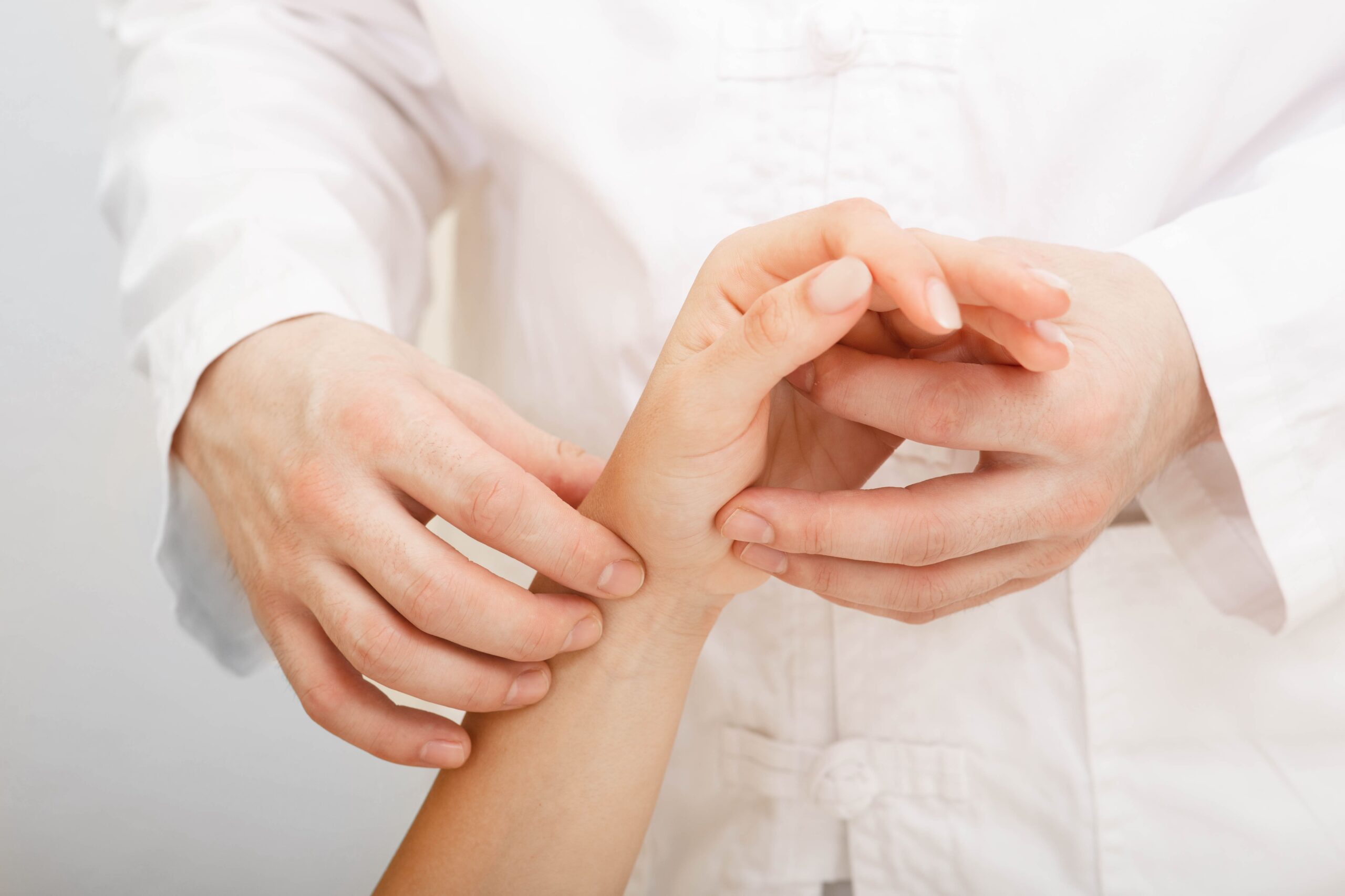
Standard X-rays may miss osteoporotic hand and wrist fractures because of poor bone contrast with surrounding soft tissues. The trabecular bone appears almost transparent on radiographs, making hairline fractures and impacted injuries difficult to identify. Hand surgeons often order multiple views, including oblique projections and comparison films of the uninjured side to detect subtle abnormalities.
CT scanning provides detailed visualisation of fractures in osteoporotic bones, revealing the extent of comminution (bone fragmentation) and articular surface involvement. Three-dimensional reconstructions help surgeons plan fixation strategies by showing the exact positions and sizes of fragments. The scan takes 5-10 minutes and requires the hand to remain still in a specific position within the scanner.
MRI evaluation becomes necessary when ligament injuries accompany fractures, which occur frequently in osteoporotic wrists. The scapholunate and lunotriquetral ligaments often tear during low-energy falls due to abnormal force distribution through weakened bones. MRI sequences optimised for bone marrow oedema also detect occult fractures invisible on standard imaging.
Bone density testing of the contralateral wrist provides essential information for treatment planning. Severe osteoporosis requires modified surgical techniques and extended healing times. The one-third radius site measurement correlates strongly with fracture risk and helps predict implant failure rates.
Modified Surgical Approaches
Osteoporotic bone presents unique surgical challenges requiring specialised techniques and implants. Standard screws achieve poor purchase in porous bone. Surgeons must modify their approach to use locking plate technology, in which screws thread directly into the plate, creating a fixed-angle construct that doesn’t rely solely on bone quality for stability.
Volar locking plates have transformed the treatment of distal radius fractures in osteoporotic patients. The plate sits on the palm side of the wrist, avoiding the extensor tendons while providing support to the articular surface. Screws placed in multiple directions create a scaffold that maintains reduction even in severely comminuted fractures. The subchondral support prevents late collapse, a common complication when treating osteoporotic fractures with cast immobilisation alone.
⚠️ Important Note
Augmentation with bone cement or synthetic bone substitutes may be necessary during surgery when screw purchase remains inadequate despite the use of locking technology.
External fixation is an alternative when internal fixation isn’t feasible due to severe comminution or soft-tissue compromise. The fixator spans the fracture site with pins placed in the metacarpals and radius shaft, maintaining length and alignment while the fracture heals. Modern low-profile fixators allow early finger motion, preventing stiffness that commonly develops during prolonged immobilisation.
Bone grafting requirements increase significantly in osteoporotic fracture surgery. Autograft from the iliac crest provides both structural support and biological healing factors, though concerns about morbidity at the harvest site exist in elderly patients. Allograft and synthetic substitutes offer alternatives that avoid donor-site pain, though incorporation times may be longer in osteoporotic bone.
Conservative Management Strategies
Non-surgical treatment remains appropriate for stable, minimally displaced fractures despite osteoporosis. Custom thermoplastic splints provide support and conform to the patient’s anatomy while allowing adjustment as swelling resolves. The splint extends from below the elbow to the metacarpal heads, immobilising the wrist while permitting finger motion.
Serial radiographs at days 7, 14, and 21 detect early displacement that occurs more frequently in osteoporotic fractures. Even minimal motion at the fracture site can lead to progressive deformity requiring surgical intervention. Weekly clinical examinations assess pain levels, swelling patterns, and early signs of complications like complex regional pain syndrome.
Physical therapy begins immediately for non-immobilised joints to prevent stiffness and maintain strength. Shoulder pendulum exercises, elbow range-of-motion exercises, and finger tendon gliding exercises may continue throughout the immobilisation period. Osteoporotic patients lose muscle mass rapidly during immobilisation, making early mobilisation protocols necessary.
Cast complications occur more frequently in osteoporotic patients due to fragile skin and poor circulation. Pressure sores can develop if the cast is too tight, while loose casts allow fracture displacement. Bi-valve casts that can be temporarily removed for skin inspection provide an alternative for high-risk patients.
Rehabilitation Considerations
Osteoporotic bones require 12-16 weeks for basic healing compared to 6-8 weeks in healthy bone. Rehabilitation protocols must account for this extended timeline while balancing the need to prevent stiffness against the risk of refracture. Hand therapy begins with gentle active range-of-motion exercises, progressing to passive stretching only after radiographic confirmation of solid union.
Strengthening exercises start with isometric contractions that don’t stress the healing fracture site. Therapy putty exercises begin at the softest resistance level, with progression occurring over months rather than weeks. Grip strength typically reaches a reduced level compared to the uninjured side even after complete healing, requiring permanent activity modifications.
✅ Quick Tip
Water-based therapy provides resistance training with reduced impact forces, making it suitable for osteoporotic patients beginning strengthening exercises.
Joint protection education becomes essential for preventing future injuries. Patients learn to use both hands for lifting, avoid sudden twisting motions, and modify daily activities to reduce fracture risk. Adaptive equipment, such as jar openers, ergonomic tools, and padded grips, reduces stress on vulnerable bones while maintaining independence.
Fall prevention strategies specifically addressing hand and wrist protection include teaching patients to fall onto their forearms rather than outstretched hands when possible. Home modifications, such as removing throw rugs, installing grab bars, and improving lighting, reduce fall risk. Balance training and tai chi programs show particular benefit for reducing fall-related wrist fractures.
Long-term Complications
Malunion occurs when osteoporotic fractures heal in suboptimal positions, leading to permanent deformity and functional limitations. The distal radius may heal with excessive dorsal angulation, leading to wrist extension weakness and pain. Corrective osteotomy becomes technically challenging in osteoporotic bone, often requiring extensive bone grafting and prolonged recovery periods.
Post-traumatic arthritis develops earlier and progresses faster in osteoporotic fracture patients. Cartilage damage from the initial injury, combined with altered joint mechanics from even minor malunion, can accelerate degenerative changes. Radiocarpal and midcarpal arthritis cause progressive pain and stiffness, eventually requiring fusion or arthroplasty procedures.
Tendon ruptures represent a delayed complication specific to osteoporotic wrist fractures. The extensor pollicis longus tendon can rupture in some distal radius fractures, occurring weeks to months after the initial injury. Sharp bone edges or prominent hardware irritate the tendon until it fails, requiring tendon transfer surgery to restore thumb extension.
Complex regional pain syndrome affects osteoporotic fracture patients more frequently than the general population. The condition causes severe burning pain, swelling, and skin changes disproportionate to the original injury. Early recognition and treatment with hand therapy, medications, and sometimes nerve blocks may help prevent permanent disability.
What Our Hand Surgeon Says
The intersection of osteoporosis and hand injuries requires thinking beyond standard fracture treatment protocols. Each patient’s bone quality directly influences surgical planning—what works for a patient with healthy bones often fails in patients with osteoporosis. Pre-contoured locking plates with multiple screw options may be used to achieve stable fixation in poor-quality bone.
Recovery expectations must be adjusted from the outset. While younger patients might return to complete activities in a few months, osteoporotic patients may need significantly longer to fully heal. This extended timeline can be challenging for active seniors, but pushing too hard, too soon, can lead to a refracture or hardware failure.
Successful outcomes involve treating the whole patient, not just the fracture. Optimising vitamin D levels, allowing adequate calcium intake, and coordinating with endocrinologists for osteoporosis medication management all contribute to better healing. Hand therapy must be gentle yet consistent, respecting the bone’s fragility while preventing debilitating stiffness.
Putting This Into Practice
- Discuss bone density testing with your hand surgeon if you’re over 65 or have risk factors for osteoporosis, particularly focusing on the distal radius measurement, which may help assess wrist fracture risk.
- Modify your home environment by installing bathroom grab bars, securing loose carpets, and ensuring adequate lighting in hallways and stairs to help prevent falls.
- Consider balance exercises such as standing on one foot or walking heel-to-toe, which may help improve stability and reduce fall risk. Consult a hand surgeon for appropriate exercise recommendations.
- Learn proper lifting techniques using your legs and core rather than putting strain on your wrists and hands.
- Consider wearing wrist guards during activities with fall risk, such as walking on ice or participating in exercise classes.
When to Seek Professional Help
- Wrist or hand pain following a fall, even if you can still move your fingers
- Swelling that doesn’t improve with ice and elevation after 48 hours
- Visible deformity or abnormal positioning of the wrist or fingers
- Numbness or tingling in the fingers that persists or worsens
- Inability to grip objects or perform daily activities due to hand weakness
- Grinding or clicking sensations in the wrist during movement
- Previous wrist fracture with new onset of pain or functional decline
Commonly Asked Questions
How long does it take for a wrist fracture to heal when you have osteoporosis?
Osteoporotic wrist fractures require 12-16 weeks for initial bone healing, compared to 6-8 weeks in healthy bone. Complete recovery, including return of strength and function, typically takes 6-9 months. Regular X-rays monitor healing progress, and your hand surgeon may adjust activity restrictions based on how well the bone is consolidating.
Can I prevent hand and wrist fractures if I already have osteoporosis?
While you can’t eliminate fracture risk entirely, several strategies can reduce it. Bisphosphonate medications improve bone density over time, vitamin D supplementation enhances calcium absorption, and targeted exercises strengthen the muscles supporting your wrists. Occupational therapy teaches joint protection techniques and provides adaptive equipment to minimise stress on fragile bones during daily activities.
Will I need surgery for every wrist fracture if I have osteoporosis?
Surgery isn’t automatic for osteoporotic wrist fractures. Stable, well-aligned fractures often heal successfully with casting or splinting. However, osteoporotic bones tend to collapse or shift position more readily than healthy bones, making surgery more likely if the fracture involves the joint surface or shows any instability on initial X-rays.
What’s different about hand surgery in patients with osteoporosis?
Surgeons use specialised implants designed for poor bone quality, including locking plates where screws thread into the plate itself rather than relying solely on bone grip. Surgery may take longer because fragile bone must be handled carefully, and surgeons often use bone grafts or cement to augment fixation. Post-operative protocols proceed more slowly with extended protection periods.
Can osteoporosis medication help my fracture heal faster?
Certain osteoporosis medications, particularly teriparatide (a parathyroid hormone analogue), may accelerate fracture healing by stimulating new bone formation. However, bisphosphonates, the most common osteoporosis drugs, need to be temporarily discontinued during early fracture healing as they may slow the initial repair process. Your hand surgeon can coordinate with your primary physician to optimise medication timing.
Next Steps

Don't Let Hand Pain Affect Your Life
Restore Function and Improve Your Quality of Life
Our clinic provides a comprehensive, one-stop service for diagnosis and treatment.
Make An EnquiryOsteoporotic hand injuries require specialised treatment approaches with extended healing timelines and modified surgical techniques. Early intervention and appropriate bone health management optimise outcomes and prevent complications.
If you are experiencing persistent hand or wrist pain, weakness with gripping, or have sustained a recent fall with ongoing symptoms, consult a hand surgeon for comprehensive evaluation and treatment.