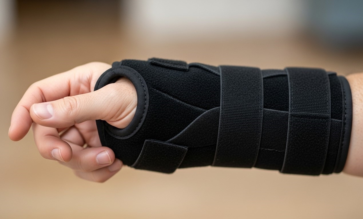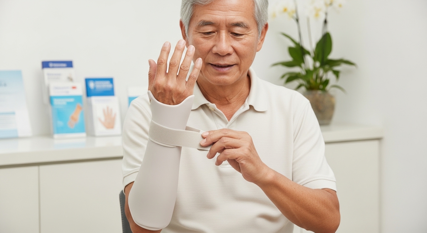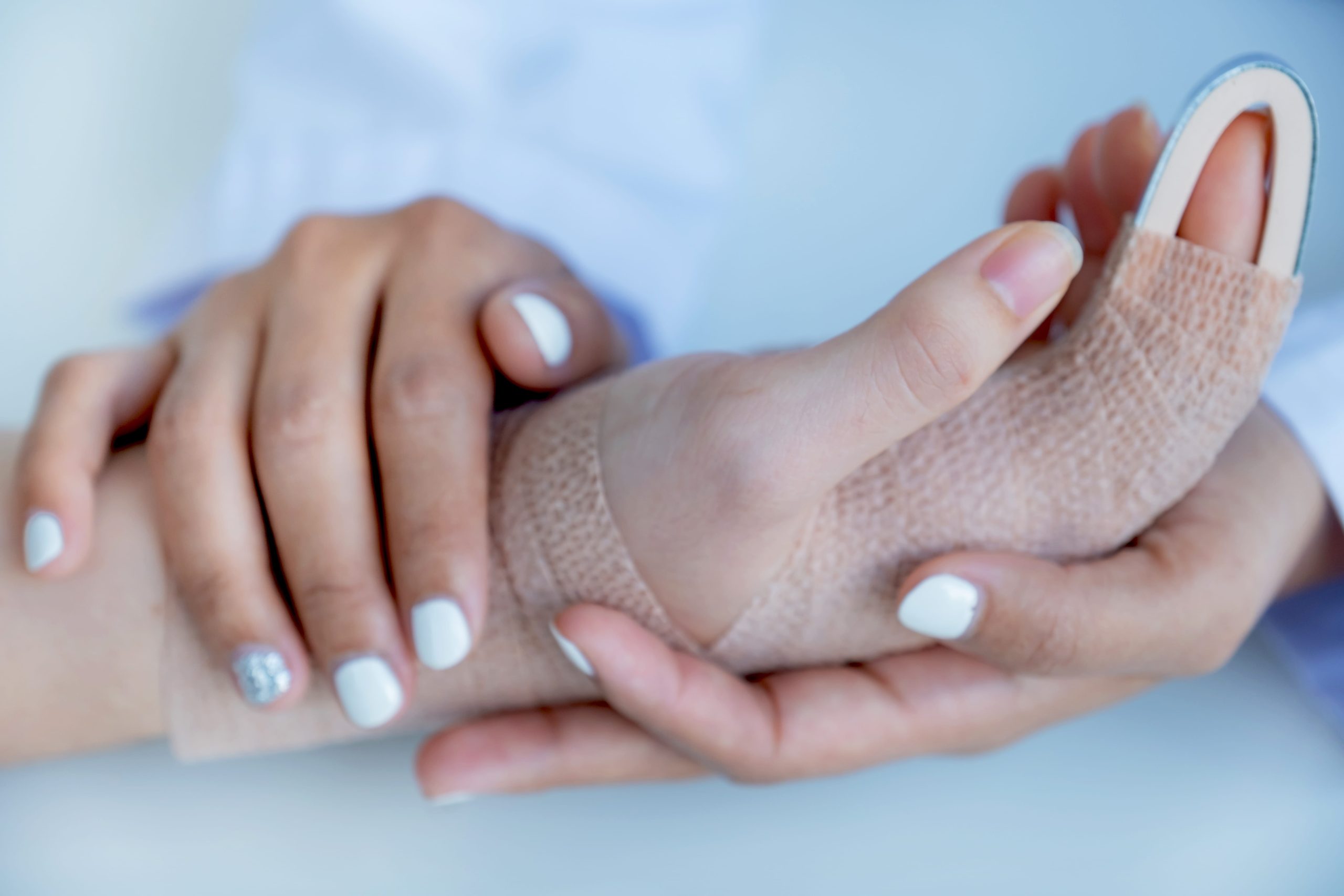
A finger that won’t straighten properly after a ball strike or fall may involve more than simple bruising. The volar plate, a thick ligament on the palm side of each finger joint, absorbs impact forces that would otherwise dislocate the joint, and when it tears or pulls away bone fragments, the injury requires different treatment than a typical jammed finger.
The proximal interphalangeal (PIP) joint, the middle knuckle of each finger, sustains these injuries most frequently, typically during catching sports, falls onto outstretched hands, or industrial accidents that force the joint into hyperextension. Recognising a volar plate injury early and distinguishing pure hyperextension from rotational forces that may also involve the collateral ligaments prevents the chronic stiffness and swan neck deformity that can develop when these injuries go untreated.
Anatomy of the Volar Plate
The volar plate consists of fibrocartilage that attaches firmly to the middle phalanx and loosely to the proximal phalanx through flexible checkrein ligaments. This asymmetric attachment allows the finger to flex fully while limiting hyperextension. The exact degree of normal passive hyperextension varies between individuals, which is why clinicians compare the injured finger to the same finger on the uninjured hand rather than relying on a fixed reference angle.
Blood supply enters primarily from the sides, so the central portion heals more slowly. The plate also serves as a gliding surface for flexor tendons, explaining why adhesions between these structures cause post-injury stiffness.
Adjacent structures compound injury complexity. The collateral ligaments attach alongside the volar plate, and the flexor tendon sheath runs directly beneath it. Swelling in this confined space creates pressure that restricts motion even after the primary injury heals.
How Volar Plate Injuries Occur
Hyperextension injuries follow a predictable pattern. The fingertip catches on a surface, a basketball, the ground, a piece of machinery, and bends backwards past its normal range. The volar plate resists this motion until tissue failure occurs.
- Type I injuries involve stretching or partial tearing without macroscopic instability. The joint remains aligned, and the finger functions with localised pain.
- Type II injuries produce complete tears with or without small bone avulsions. The joint may subluxate momentarily but reduces spontaneously. These represent a significant portion of volar plate injuries managed clinically.
- Type III injuries combine volar plate disruption with fracture-dislocations. The middle phalanx displaces dorsally and remains unstable, often requiring surgical evaluation.
Contact sports commonly generate Type I and II injuries. Rugby, basketball, netball, and volleyball players experience jammed fingers regularly, with a subset sustaining volar plate damage. Capsular injuries from hyperextension are also seen in combat sports, and climbing can produce similar injuries when fingers catch during falls.
Distinguishing Volar Plate Injuries from Simple Jammed Fingers
Both conditions cause immediate pain and swelling at the PIP joint. The differences emerge over the following days.
- Simple jammed fingers typically improve steadily. Swelling generally decreases, motion returns, and function commonly normalises within two to three weeks under standard care. The joint remains stable throughout its range of motion.
- Volar plate injuries may plateau or worsen without proper management. Swelling can persist around the joint, and a specific tender point exists on the palm side of the PIP joint. The finger may hyperextend passively more than the same finger on the opposite hand, indicating potential structural damage.
Testing lateral stability reveals collateral ligament involvement. With the joint slightly flexed, side-to-side stress should produce minimal movement. Laxity indicates a combined injury requiring comprehensive rehabilitation planning.
The “pseudoboutonnière” position, slight flexion at the PIP joint with difficulty achieving full extension, suggests significant volar plate damage. This differs from a true boutonnière deformity, where the central slip of the extensor tendon ruptures.
Diagnostic Evaluation
Clinical examination provides substantial information before imaging. The hand surgeon assesses swelling location, point tenderness, active and passive range of motion, and joint stability in multiple planes.
X-rays remain the first-line imaging study. Standard lateral views may show small avulsion fractures at the volar plate attachment site. The size of these fragments influences treatment decisions; fragments involving more than 30-40% of the articular surface suggest instability requiring surgical consideration.
Dorsal subluxation of the middle phalanx on lateral x-ray indicates fracture-dislocation. Even subtle dorsal translation changes the treatment approach significantly.
Ultrasound visualises soft tissue damage that X-rays miss. Dynamic scanning while moving the finger reveals instability patterns and helps distinguish partial from complete tears.
MRI serves specific situations: diagnostic uncertainty, suspected combined injuries, or pre-operative planning for complex cases. The expense and time required make MRI unnecessary for straightforward presentations.
Non-Surgical Treatment Approaches
Most volar plate injuries heal with appropriate conservative management. The treatment timeline spans six to eight weeks, with technique details affecting outcomes substantially.
Did You Know?
The volar plate heals in a shortened position when immobilised in full extension. This creates permanent hyperextension laxity, the opposite of the desired outcome. Proper splinting positions the joint in slight flexion to allow healing at appropriate tension.
Extension block splinting forms the foundation of non-surgical treatment. A dorsal aluminium splint prevents the last 20-30 degrees of extension while allowing full flexion. This position protects the healing volar plate while maintaining tendon gliding and preventing stiffness.
The extension block decreases progressively over four to six weeks. Weekly adjustments of approximately 10 degrees eventually permit full extension as tissue strength returns.
Active motion within the protected range begins immediately. Flexion exercises maintain tendon excursion and joint nutrition. Patients perform these exercises multiple times daily, removing the splint only for supervised movement initially.
Oedema control accelerates recovery. Elevation, compression wraps, and contrast baths reduce swelling that otherwise limits motion. Persistent oedema correlates with increased scarring and stiffness.
Surgical Treatment Options
Surgery addresses unstable fracture-dislocations, large avulsion fragments, and injuries that fail conservative treatment after an adequate trial period.
Extension block pinning treats fracture-dislocations where closed reduction achieves joint congruity. A wire placed dorsally prevents the middle phalanx from subluxating while early motion proceeds. The wire is removed in the clinic after three to four weeks.
Open reduction and internal fixation applies to large articular fragments. Screws or plates restore joint surface anatomy when fragment size permits fixation. Post-operative rehabilitation follows similar principles to conservative treatment.
Volar plate arthroplasty addresses chronic injuries where the volar plate has healed in an incompetent position. The surgeon advances the scarred plate to restore stability, accepting some flexion limitation in exchange for preventing hyperextension.
Hemi-hamate reconstruction salvages severely comminuted fracture-dislocations. A bone graft from the hamate in the wrist replaces the destroyed articular surface. This procedure offers options when other methods cannot restore joint function.
Recovery and Rehabilitation Timeline
Rehabilitation intensity determines functional outcomes as much as the initial treatment choice. The PIP joint can develop stiffness rapidly, and recovering motion requires sustained, guided effort.
- Weeks 1-2: Oedema control and protected active flexion within the splint parameters. Local pain typically begins to decrease gradually during this window.
- Weeks 3-4: Progressive extension block adjustment under clinical guidance. Introduction of gentle active-assisted flexion stretching may be considered if active movement plateaus.
- Weeks 5-6: Continued extension block reduction toward neutral. Guided strengthening exercises commonly begin with specialised putty or soft therapy balls.
- Weeks 7-8: Gradual weaning from the splint during daily activities. Night splinting may continue if an extension lag persists.
- Weeks 9-12: Potential return to sport or more demanding activities with buddy taping support. Full functional recovery commonly requires three to four months for athletes requiring high grip strength.
Important Note: PIP joint stiffness remaining after three months becomes increasingly complex to manage. Early hand therapy collaboration for patients who are not progressing as expected supports a more predictable recovery trajectory.
Potential Complications
Flexion contracture represents the most common complication. The joint loses terminal extension, settling into a slightly bent position. Mild contractures under 20 degrees rarely limit function, while severe contractures significantly impair hand use.
Swan neck deformity develops when volar plate incompetence allows chronic hyperextension. The extensor mechanism shifts dorsally, creating a characteristic posture with PIP hyperextension and DIP flexion. Prevention through adequate initial treatment remains easier than surgical correction.
Post-traumatic arthritis follows injuries involving articular cartilage damage. Incongruent joint surfaces or prolonged instability accelerate cartilage wear. Symptoms may not appear for years after the initial injury.
Chronic instability results from inadequate healing. The finger hyperextends easily, catches during gripping activities, and may subluxate with minimal force. Reconstruction surgery can address this, but it involves a longer recovery than primary treatment.
When to Seek Professional Help
- The finger remains swollen two weeks after the injury without improvement
- Inability to fully straighten the finger at the middle joint
- Visible deformity or deviation of the finger
- Instability or “giving way” sensation during gripping
- Catching or locking during finger movement
- Pain prevents return to normal activities after three weeks
- Hyperextension of the PIP joint compared to the opposite hand
Commonly Asked Questions
How long should I buddy tape my finger after a volar plate injury?
Buddy taping commonly continues for four to six weeks after weaning from a formal splint, particularly during sports or manual work. The adjacent finger provides dynamic stability while final tissue strengthening occurs.
Can I still play sports during recovery?
Participation in non-contact activities may often resume once localised pain permits, frequently by week three or four. High-impact contact sports and activities requiring a strong grip typically require waiting until weeks eight to twelve, depending on tissue healing progress.
Will my finger ever straighten completely again?
Many patients regain functional extension, though subtle differences compared to the uninjured finger are frequently observed clinically. A residual loss of five to ten degrees of terminal extension rarely impacts general daily activities. Patient compliance with targeted rehabilitation and splinting timelines heavily influences individual recovery.
What’s the difference between a jammed finger and a volar plate injury?
A jammed finger is a broad description of symptoms, such as localised pain and swelling after an impact, rather than a specific medical diagnosis. A volar plate injury represents one specific structural cause of these symptoms, alongside fractures, collateral ligament sprains, or deep tissue contusions. An accurate diagnosis is essential to determine the appropriate care path.
Do all volar plate injuries need surgery?
The majority of these injuries respond well to structured splinting and physical therapy alone. Surgical intervention typically becomes necessary when structural fracture fragments compromise joint alignment, when the joint cannot be stabilised conservatively, or when a dedicated trial of non-surgical management does not restore baseline stability.
Next Steps
Most volar plate injuries are treatable with extension block splinting when diagnosed within the first few weeks, before scar tissue and contracture develop. Splinting must position the joint in slight flexion, not full extension, to allow the volar plate to heal at correct tension. Injuries involving joint instability or articular fragments greater than 30 to 40% of the joint surface require surgical evaluation.
If you are experiencing persistent finger swelling, difficulty straightening your finger at the middle knuckle, or joint instability or hyperextension after an injury, consult our hand surgeon for an evaluation and targeted treatment plan.