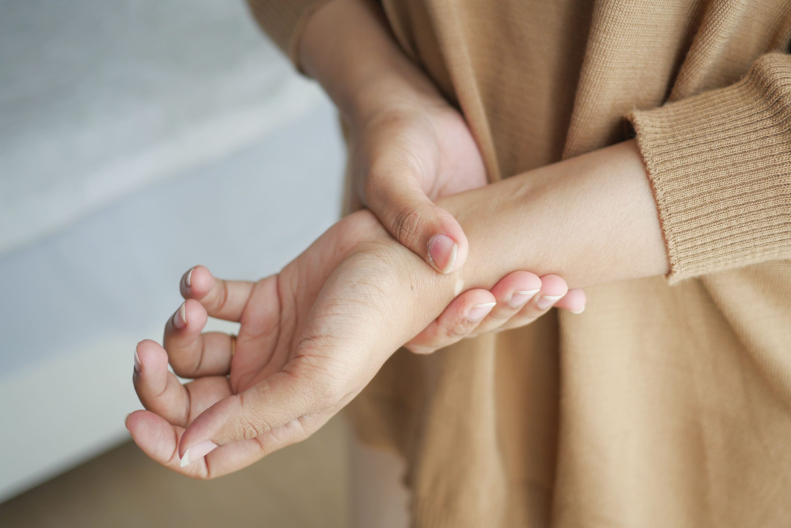
The triangular fibrocartilage complex (TFCC) acts as a shock absorber between your ulna bone and the small carpal bones on the pinky side of your wrist. This structure, composed of cartilage, ligaments, and tendons, stabilises your wrist during gripping and rotation movements. TFCC injuries cause pain on the ulnar (pinky) side of the wrist, particularly during twisting motions like opening jars or turning doorknobs.
TFCC tears occur through two main mechanisms: acute trauma from falling on an outstretched hand or repetitive stress from activities requiring frequent wrist rotation. Athletes in racquet sports, gymnasts, and manual labourers facea higher risk due to repetitive loading patterns. The injury spectrum ranges from minor strains that heal within weeks to complete tears requiring surgical repair.
Anatomy of the TFCC
The TFCC sits between the end of your ulna bone and two small wrist bones called the lunate and triquetrum. This triangular structure measures approximately 5mm thick at its centre and consists of several interconnected components working together for wrist stability.
The central articular disc, made of fibrocartilage similar to knee meniscus tissue, absorbs compressive forces during weight-bearing activities. The radioulnar ligaments, both dorsal and palmar portions, prevent excessive movement between the radius and ulna bones during forearm rotation. The ulnocarpal ligaments connect the ulna to the carpal bones, while the extensor carpi ulnaris tendon sheath provides additional support along the ulnar border.
Blood supply to the TFCC varies significantly across its structure. The peripheral 15-20% receives rich vascularisation from branches of the ulnar artery, enabling natural healing potential. The central portion remains avascular, similar to the inner knee meniscus, explaining why central tears rarely heal without intervention. This vascular pattern directly influences treatment decisions and recovery expectations.
During normal wrist function, the TFCC bears approximately 20% of the axial load across the wrist joint. This percentage increases with ulnar deviation and pronation, explaining why these movements often trigger pain in TFCC injuries.
Recognising TFCC Injury Symptoms
Ulnar-sided wrist pain defines the hallmark symptom of TFCC injury, typically worsening with specific movements and positions. The pain localises to the depression between the ulnar styloid and flexor carpi ulnaris tendon, an area clinicians call the “fovea sign” location.
Gripping activities intensify discomfort, particularly when combined with ulnar deviation or pronation. Patients frequently report difficulty with tasks like using scissors, turning keys, or pushing up from a chair using the affected hand. The pain character varies from sharp catching sensations during movement to deep aching after prolonged use.
Mechanical symptoms develop in many TFCC tear cases. Clicking or popping sounds during wrist rotation indicate unstable tear fragments catching between joint surfaces. Some patients experience a sensation of the wrist “giving way” during load-bearing activities, reflecting compromised stability from ligamentous disruption.
Swelling appears primarily along the ulnar wrist border, though significant visible swelling remains uncommon except in acute traumatic cases. Morning stiffness lasting 15-30 minutes often accompanies chronic TFCC pathology, improving with gentle movement but returning after periods of immobility.
Range of motion limitations manifest subtly in TFCC injuries. Pronation and supination often decrease by 10-15 degrees compared to the unaffected side. Ulnar deviation typically produces pain at the end range, while radial deviation remains relatively comfortable. Grip strength measurements show a 20-30% reduction compared to the contralateral hand, with greater deficits during sustained gripping tasks.
Diagnostic Approach
Physical examination for TFCC injury employs specific provocative tests that reproduce symptoms by stressing the damaged structures. The fovea sign test involves pressing into the soft spot between the ulnar styloid and flexor carpi ulnaris tendon – tenderness here shows 95% sensitivity for TFCC pathology.
The ulnocarpal stress test combines axial loading with ulnar deviation and rotation. The examiner applies compression through the wrist while moving it through pronation and supination arcs. Pain reproduction, particularly with a palpable click, indicates TFCC tear or instability. The press test requires patients to push themselves up from a chair using their wrists in extension – ulnar-sided pain during this manoeuvre suggests TFCC involvement.
Plain radiographs provide initial screening information despite not visualising the TFCC directly. Ulnar variance measurements determine whether the ulna sits level with, shorter than, or longer than the radius at the wrist joint. Positive ulnar variance (ulna longer than radius by >2mm) increases TFCC injury risk and influences treatment planning. Radiographs also exclude fractures, arthritis, or other bony abnormalities mimicking TFCC symptoms.
MRI arthrography achieves 85-95% accuracy for tear detection. Contrast injection into the radiocarpal joint enhances visualisation of tear patterns and ligamentous disruption. High-resolution 3-Tesla MRI protocols without contrast provide reasonable diagnostic accuracy while avoiding injection procedures. MRI findings classify tears by Palmer classification: Class 1 (traumatic) tears show distinct patterns based on location, while Class 2 (degenerative) tears demonstrate fraying and thinning.
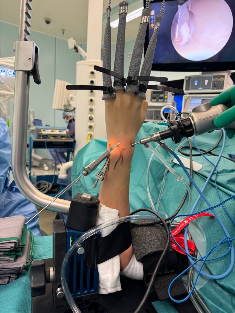
Diagnostic wrist arthroscopy provides a definitive assessment when MRI imaging remains inconclusive or surgical planning requires detailed evaluation. The arthroscope visualises tear characteristics, stability testing, and concurrent pathology not apparent on MRI. Arthroscopic findings guide immediate treatment decisions, as many tears can be debrided or repaired during the same procedure.
Treatment Strategies
Conservative Management
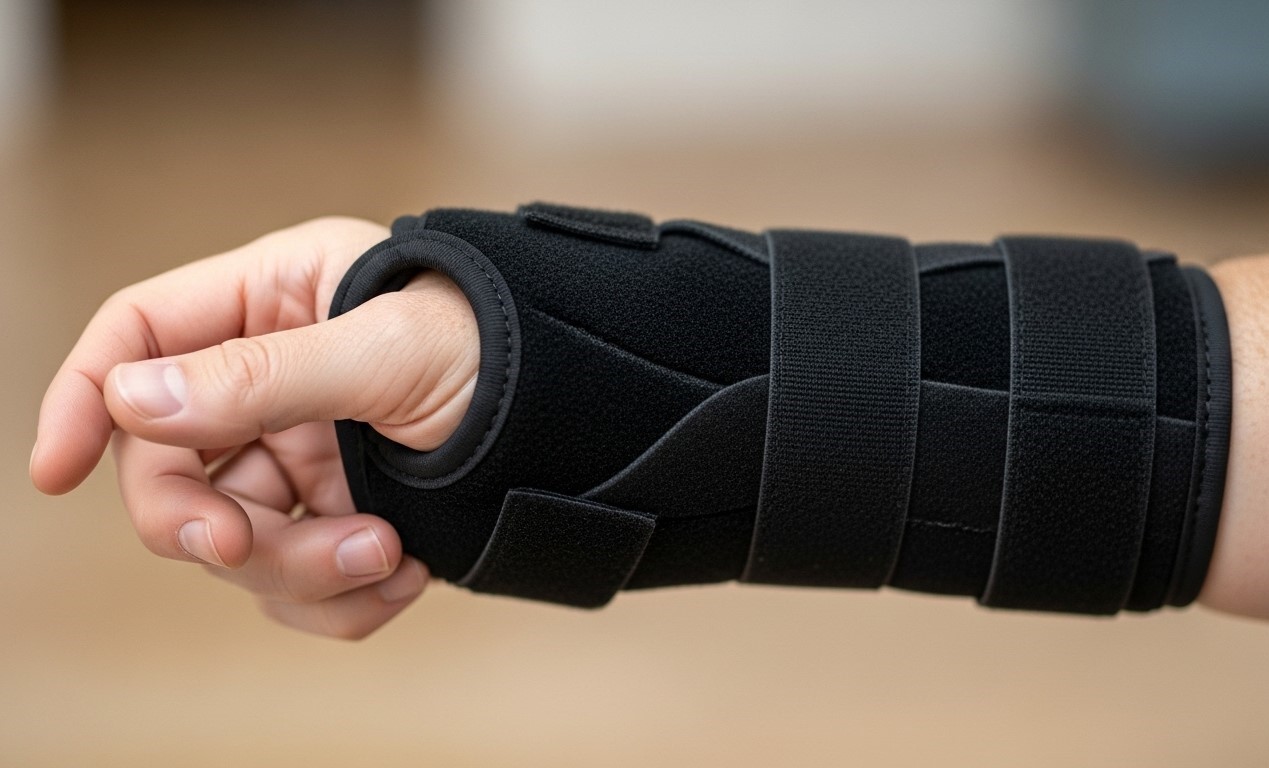
Initial treatment for TFCC injuries follows structured conservative protocols lasting 6-12 weeks. Immobilisation using a wrist splint or cast maintains the wrist in slight extension and neutral rotation, allowing inflammation to subside while preventing further tissue damage. Removable splints permit hygiene and gentle exercises while maintaining protection during daily activities.
Activity modification eliminates provocative movements during the healing phase. Patients avoid repetitive gripping, twisting motions, and weight-bearing through the affected wrist. Ergonomic adjustments at work stations reduce cumulative stress – vertical computer mice, padded wrist supports, and modified tool grips minimise ulnar loading patterns.
Hand therapy begins once acute pain subsides, typically after 2-3 weeks of immobilisation. Therapists employ specific techniques targeting TFCC stability without excessive stress. Isometric strengthening in neutral wrist positions progresses to controlled eccentric exercises. Proprioceptive training using therapy putty and weighted balls restores neuromuscular control. Manual therapy techniques address compensatory patterns in the forearm and elbow developed during the injury period.
Corticosteroid injections provide diagnostic and therapeutic benefits for persistent symptoms. MRI-guided injection ensures accurate placement into the ulnocarpal joint space or directly adjacent to the TFCC. Pain relief lasting several weeks confirms TFCC-related symptoms while potentially promoting healing through inflammation reduction. Injection frequency limited to 2-3 per year prevents potential cartilage deterioration.
Surgical Interventions
Arthroscopic debridement addresses central TFCC tears lacking healing potential due to poor vascularity. The procedure removes unstable tear fragments, causing mechanical symptoms while preserving stable peripheral tissue. Radiofrequency ablation smooths frayed edges, eliminating catching sensations during movement. Recovery requires 4-6 weeks of protected motion followed by gradual strengthening.
Peripheral TFCC repairs capitalise on the vascular zone’s healing capacity. Arthroscopic techniques use specialised suture devices to reattach torn edges to the joint capsule or foveal insertion. Outside-in, inside-out, or all-inside repair technique selection depends on tear location and surgeon preference. Post-operative protocols mandate 6 weeks of immobilisation in slight supination, followed by progressive mobilisation over 3 months.
Open repair becomes necessary for complex tears involving foveal detachment or concurrent ligament reconstruction. The surgeon accesses the TFCC through a dorsal incision, directly visualising and repairing all damaged structures. Bone tunnels or suture anchors secure the TFCC to its anatomic footprint on the ulnar fovea. Associated procedures like ulnar shortening osteotomy address positive ulnar variance contributing to TFCC overload.
Ulnar shortening osteotomy treats TFCC injuries associated with ulnocarpal impaction syndrome. The procedure removes 2-4mm of ulnar shaft, reducing mechanical stress on the TFCC. Plate fixation maintains alignment during bone healing over 8-12 weeks. This procedure shows good outcomes for degenerative TFCC tears with positive ulnar variance exceeding 2mm.
Recovery Timeline and Rehabilitation
Week 1-2 post-injury or surgery focuses on inflammation control and protection. The wrist remains immobilised in a custom splint, maintaining 15-20 degrees extension. Finger movements prevent stiffness while avoiding wrist motion. Elevation and ice application for 15-minute intervals reduce swelling.
Week 3-6 introduces gentle active range of motion exercises within pain-free ranges. Tendon gliding exercises prevent adhesions while respecting healing tissues. Scar massage begins for surgical patients once incisions heal completely. Therapists monitor for signs of complex regional pain syndrome, addressing any abnormal pain patterns immediately.
Week 7-12 progresses to strengthening exercises using resistance bands and light weights. Eccentric strengthening protocols target the extensor carpi ulnaris and flexor carpi ulnaris muscles supporting the ulnar wrist. Proprioceptive exercises on unstable surfaces challenge dynamic stability. Functional activities gradually reintroduce rotational movements and light gripping tasks.
Month 3-6 advances toward full activity resumption. Sport-specific training for athletes includes progressive loading patterns mimicking their activity demands. Manual workers practice job-specific tasks with increasing duration and intensity. Objective testing using dynamometry and functional assessments confirms readiness for unrestricted activity.
💡 Did You Know?
The TFCC experiences maximum stress during the “dart thrower’s motion” – a diagonal movement from radial extension to ulnar flexion. This motion pattern explains why many patients develop compensatory movement strategies avoiding this specific arc.
Complications and Long-term Outlook
Untreated TFCC injuries lead to progressive ulnocarpal instability and degenerative changes. Chronic inflammation causes synovitis, producing persistent swelling and stiffness. Altered wrist mechanics from TFCC insufficiency accelerate cartilage wear, potentially developing into ulnocarpal arthritis within several years.
Failed conservative treatment occurs in some complete tears, particularly those involving peripheral detachment or associated instability. Persistent mechanical symptoms despite adequate conservative management indicate structural failure requiring surgical intervention. Delayed surgery after failed conservative treatment shows comparable outcomes to early surgical intervention.
Post-surgical complications remain uncommon but require prompt recognition. Infection rates stay below 1% with proper sterile technique and perioperative antibiotics. Nerve irritation, particularly of the dorsal sensory branch of the ulnar nerve, causes temporary numbness along the ulnar hand border. Stiffness from prolonged immobilisation responds to dedicated therapy protocols emphasising progressive mobilisation.
⚠️ Important Note
TFCC re-tears following surgical repair occur most frequently during the first 3 months post-operatively. Strict adherence to post-operative protocols and avoiding premature loading significantly reduces re-injury risk.
Putting This Into Practice
- Document your pain patterns in a symptom diary, noting specific activities that trigger discomfort and positions that provide relief – this information guides your specialist’s treatment planning
- Modify your workspace ergonomics immediately by adjusting keyboard height to maintain a neutral wrist position and using padded wrist supports during computer work
- Begin gentle tendon gliding exercises (making a fist, then straightening fingers, then bending at knuckles only) to maintain flexibility without stressing the TFCC
- Apply ice wrapped in a thin towel to the ulnar wrist for 15-minute sessions when pain increases after activity
- Practice protective techniques during daily activities, such as pushing doors open with your shoulder instead of your hand and using both hands for lifting tasks
When to Seek Professional Help
- Ulnar-sided wrist pain persisting beyond one week despite rest and activity modification
- Clicking or popping sensations during wrist rotation movements
- Wrist instability or feeling of “giving way” during gripping activities
- Swelling along the pinky side of the wrist that doesn’t respond to ice and elevation
- Difficulty performing daily tasks like turning doorknobs or opening jars
- Pain that wakes you at night or throbs at rest
- Numbness or tingling extending into the ring and small fingers
Commonly Asked Questions
How long does a TFCC injury take to heal without surgery?
Minor TFCC strains resolve within 4-6 weeks with proper immobilisation and activity modification. Partial tears require 8-12 weeks of conservative treatment, though healing depends on tear location and blood supply. Central tears lacking vascularity rarely heal completely without surgical intervention.
Can I continue exercising with a TFCC injury?
Lower body exercises and cardiovascular activities that don’t load the wrist remain safe during recovery. Swimming requires modification to avoid wrist extension during strokes. Weight training must eliminate exercises requiring gripping or wrist loading until cleared by your specialist.
What’s the difference between TFCC injury and wrist tendinitis?
TFCC injuries produce specific ulnar-sided pain worsening with rotation and compression, often accompanied by mechanical symptoms. Tendinitis causes diffuse pain along tendon paths, worsening with repetitive motion but improving with rest. TFCC injuries show positive provocative tests like the fovea sign, while tendinitis produces pain with resisted muscle testing.
Will I need surgery for my TFCC tear?
Peripheral tears with good blood supply often heal with conservative treatment. Central or complex tears causing persistent mechanical symptoms typically require arthroscopic intervention. Your hand specialist determines surgical necessity based on tear characteristics, symptom severity, and response to initial treatment.
How successful is TFCC surgery?
Arthroscopic debridement for central tears achieves good outcomes in many patients. Peripheral repairs show high satisfaction rates when performed within the vascular zone. Success depends on tear pattern, surgical technique, and adherence to rehabilitation protocols.
Next Steps
TFCC injuries require an accurate diagnosis to distinguish from other causes of ulnar wrist pain. Early intervention prevents progression to chronic instability or degenerative changes. Whether your injury stems from acute trauma or repetitive stress, proper evaluation guides treatment selection between conservative management and surgical options.
If you’re experiencing ulnar-sided wrist pain, clicking sensations, or grip weakness mentioned in this article, our hand specialist can provide a comprehensive evaluation and treatment options.

Don't Let Hand Pain Affect Your Life
Restore Function and Improve Your Quality of Life
Our clinic provides a comprehensive, one-stop service for diagnosis and treatment.
Make An Enquiry