Wrist fractures can damage the median, ulnar, or radial nerves through direct trauma, bone fragment pressure, or swelling-induced compression. These nerves control sensation and movement in specific hand regions – the median nerve affects the thumb through the middle finger, the ulnar nerve controls the ring and small fingers, while the radial nerve manages wrist extension and thumb movement.
Nerve injuries occur through three mechanisms during wrist fractures. Direct impact can tear or bruise nerve fibres at the moment of injury. Displaced bone fragments may compress nerves against surrounding structures. Post-injury swelling creates pressure within tight anatomical spaces, particularly the carpal tunnel, where the median nerve travels.
The severity of nerve damage after wrist fracture ranges from temporary dysfunction to complete nerve disruption requiring surgical repair. Early recognition of nerve symptoms allows timely intervention before permanent changes develop in muscles and sensory receptors.
Types of Nerve Injuries in Wrist Fractures
Neuropraxia
Neuropraxia represents the mildest form of nerve injury, where the nerve remains intact but cannot transmit signals properly. The protective myelin sheath experiences temporary dysfunction from compression or stretching. Patients experience numbness, tingling, or weakness in the affected nerve distribution that typically resolves within 6-12 weeks without intervention.
Recovery follows a predictable pattern – sensory function returns first, followed by motor strength. The nerve regenerates its myelin coating at approximately 1mm per day. During recovery, patients may experience electric shock sensations or hypersensitivity as nerve function returns.
Axonotmesis
Axonotmesis involves the disruption of nerve fibres while the surrounding connective tissue remains intact. The nerve must regenerate from the point of injury toward the fingertips at 1-3mm per day. Complete recovery requires 3-6 months, depending on the injury location – wrist-level injuries need less regeneration distance than forearm fractures.
This injury type commonly occurs from severe compression or traction during high-energy fractures. The intact nerve sheath guides regenerating fibres to their original targets. Patients experience a complete loss of function below the injury site initially, with a gradual return following the proximal-to-distal regeneration pattern.
Neurotmesis
Neurotmesis represents a complete nerve transection requiring surgical repair. Without surgery, regenerating nerve fibres cannot cross the gap or may form painful neuromas. Surgical options include direct repair for clean cuts with minimal gap, nerve grafting for larger defects, or nerve transfers using nearby donor nerves.
Timing affects outcomes significantly – repairs within 3-6 months achieve better results than delayed interventions. After this window, muscles undergo irreversible changes and sensory receptors degenerate. Even with optimal surgical repair, recovery remains incomplete with permanent sensory changes and some weakness.
Recognising Nerve Damage Symptoms
Sensory Changes
Numbness and tingling follow specific patterns based on the affected nerve. Median nerve compression causes numbness in the thumb, index, middle, and half of the ring finger. Ulnar nerve involvement affects the small finger and half of the ring finger. Radial nerve sensory loss occurs over the dorsal first web space between the thumb and index finger.
Hypersensitivity develops as nerves regenerate, causing painful responses to light touch or temperature changes. Patients describe burning, electric, or stabbing sensations triggered by normally non-painful stimuli. This hypersensitivity indicates active nerve recovery but can significantly impact daily activities and sleep quality.
Loss of protective sensation increases injury risk. Patients may burn themselves without awareness or develop wounds from prolonged pressure. Temperature discrimination disappears before light touch sensation, making hot water or cold objects particularly dangerous.
Motor Dysfunction
Median nerve motor loss prevents thumb opposition – the ability to touch the thumb tip to other fingertips. Patients cannot perform pinch grips for writing, buttoning clothes, or picking up small objects. The thenar muscles at the thumb base atrophy within weeks, creating a flattened appearance.
Ulnar nerve dysfunction causes small finger and ring finger weakness, with the inability to spread fingers apart or bring them together. The hand develops a “claw” position with hyperextension at the knuckles and flexion at the finger joints. Fine motor tasks like typing or playing instruments become impossible.
Radial nerve palsy produces “wrist drop”, where the wrist cannot extend against gravity. Patients compensate by flexing the wrist to release their grip, creating the characteristic “waiter’s tip” position when attempting to shake hands. Thumb and finger extension at the knuckle joints also fails.
Autonomic Symptoms
Nerve damage disrupts sweat production and blood flow regulation in affected areas. Dry, shiny skin develops over denervated regions, while adjacent areas may sweat excessively. The skin becomes thin and easily injured without normal nerve-regulated maintenance.
Temperature regulation fails in denervated digits, causing persistent coldness or inappropriate warming. Nail growth slows or becomes irregular. Hair growth patterns change with either excessive growth or complete loss in affected areas.
Diagnostic Evaluation
Clinical Testing
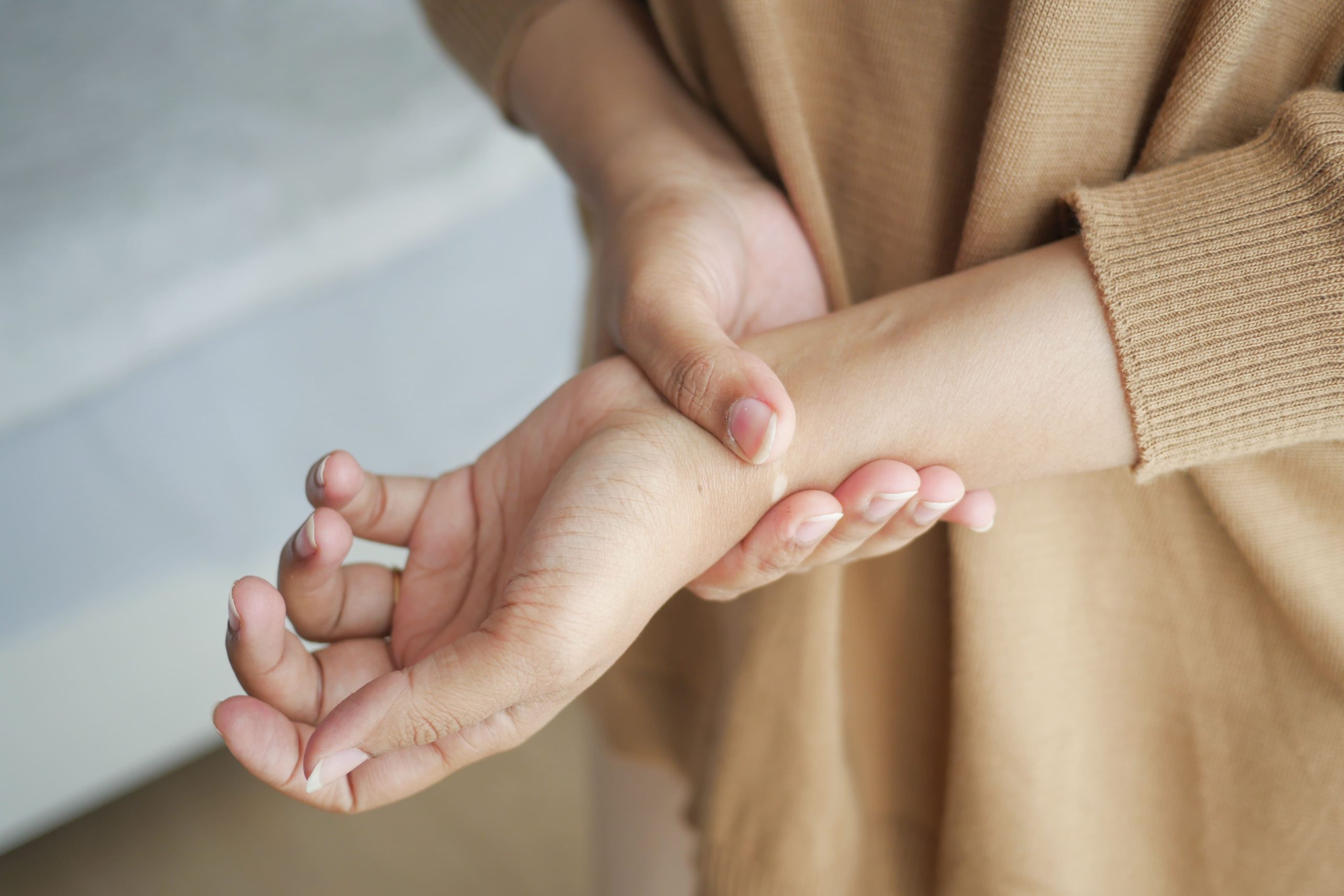
Physical examination reveals specific patterns that distinguish between nerve injuries. Tinel’s sign – tingling when tapping over the nerve – indicates the regeneration front location. A progressively advancing Tinel’s sign suggests ongoing recovery, while a stationary sign may indicate blocked regeneration.
Two-point discrimination testing measures sensory nerve function objectively. Normal fingertip discrimination ranges from 3-5mm. Values exceeding 10mm indicate significant sensory loss requiring close monitoring. Serial measurements track recovery progress.
Manual muscle testing grades strength from 0 (no contraction) to 5 (normal strength). Individual muscle testing identifies specific nerve involvement – thumb abduction tests the median nerve, finger spreading tests the ulnar nerve, and wrist extension tests radial nerve function.
Electrodiagnostic Studies
Nerve conduction studies measure electrical signal transmission speed and amplitude. Slowed conduction velocity indicates demyelination from compression. Reduced amplitude suggests axon loss requiring longer recovery. Studies performed 3-4 weeks post-injury provide baseline values for comparison.
Electromyography (EMG) examines muscle electrical activity, revealing denervation patterns. Fibrillation potentials appear 2-3 weeks after complete nerve injury. Reinnervation produces characteristic electrical patterns months before clinical strength returns. Serial EMG studies guide prognosis and treatment decisions.
Imaging Studies
MRI visualises nerve continuity, swelling, and compression sites in real-time. Dynamic scanning during wrist movement reveals nerve entrapment or tethering. MRI-guided injections deliver medication precisely to compressed nerve sites.
MRI provides detailed nerve anatomy and surrounding soft tissue evaluation. Special sequences highlight nerve oedema, scarring, or discontinuity. MRI neurography techniques trace the entire nerve path, identifying multiple compression sites.
Treatment Approaches
Conservative Management
Splinting maintains optimal wrist position, reducing nerve tension and preventing further injury. Neutral wrist positioning minimises carpal tunnel pressure on the median nerve. Night splints prevent prolonged wrist flexion during sleep, which exacerbates nerve compression.
Medications target different aspects of nerve symptoms. Gabapentin or pregabalin reduce neuropathic pain and hypersensitivity. Short-term oral steroids decrease inflammation around compressed nerves. Vitamin B complex supports nerve regeneration, though evidence remains limited.
Hand therapy begins immediately, focusing on oedema control through elevation, compression, and active motion of unaffected joints. Desensitisation techniques gradually normalise hypersensitive areas using textures progressing from soft to rough. Nerve gliding exercises prevent adhesions while maintaining nerve mobility through the available range of motion.
Surgical Interventions
Nerve decompression surgery relieves pressure from bone fragments, scar tissue, or tight anatomical tunnels. Carpal tunnel release for median nerve compression involves dividing the transverse carpal ligament through a small palmar incision. Cubital tunnel release frees the ulnar nerve at the elbow if compression occurs at multiple levels.
Nerve repair techniques depend on injury pattern and timing. Primary repair reconnects cleanly cut nerves using microscopic sutures smaller than human hair. Nerve grafting bridges gaps using expendable sensory nerves, commonly the sural nerve from the leg. Processed nerve allografts avoid donor site morbidity for gaps under 5cm.
Tendon transfers restore function when nerve recovery fails or remains incomplete after 12-18 months. Functioning muscles redirect to perform lost movements – flexor tendons can restore thumb opposition after median nerve palsy. Multiple transfers may recreate complex movements like finger extension after radial nerve injury.
Recovery Timeline and Rehabilitation
Early Phase (0-6 weeks)
Initial recovery focuses on protecting healing fractures while preventing stiffness and managing swelling. Gentle active motion of unaffected joints maintains mobility. Mirror therapy tricks the brain by watching the unaffected hand move, potentially accelerating nerve recovery.
Sensory re-education begins once fractures stabilise. Patients practice identifying textures, shapes, and temperatures with vision, then without. Localisation exercises involve touching specific points with eyes closed. These exercises maintain cortical representation of the affected area during nerve regeneration.
Intermediate Phase (6-12 weeks)
Progressive strengthening begins as fractures consolidate. Isometric exercises activate muscles without joint movement. Therapy putty exercises progress from soft to firm resistance. Functional electrical stimulation maintains muscle bulk in completely denervated muscles.
Sensory recovery typically begins during this phase, with deep pressure sensation returning before light touch. Patients practice graded motor imagery – imagining movements, recognising hand positions in photographs, then performing mirror therapy before attempting actual movement.
Late Phase (3-6 months)
Advanced strengthening uses progressive resistance and functional activities. Work simulation prepares for vocational demands. Adaptive equipment compensates for persistent deficits while maximising available function. Patients learn one-handed techniques for bilateral tasks if recovery plateaus.
💡 Did You Know?
Nerve regeneration continues for up to two years after injury, though the rate slows progressively. Younger patients typically experience faster and more complete recovery due to superior regeneration capacity and neural plasticity.
Complications and Long-term Outcomes
Chronic Regional Pain Syndrome
Complex regional pain syndrome (CRPS) develops in some patients following nerve damage after a wrist fracture. Severe burning pain disproportionate to the injury, swelling, colour changes, and abnormal sweating characterise this condition. Early recognition and aggressive therapy prevent progression to irreversible changes.
Treatment requires multidisciplinary approaches combining medications, therapy, and psychological support. Stellate ganglion blocks interrupt abnormal sympathetic signals. Spinal cord stimulation may help refractory cases. Movement-based therapies, despite pain, remain fundamental to recovery.
Permanent Deficits
Incomplete recovery leaves varying degrees of permanent dysfunction. Sensory deficits typically involve altered sensation rather than complete numbness – patients describe feeling through gloves or persistent tingling. Protective sensation usually returns even when fine discrimination remains impaired.
Motor recovery plateaus by 18-24 months post-injury. Residual weakness affects grip strength and endurance more than crude power. Intrinsic muscle atrophy may persist despite nerve recovery if reinnervation occurs after irreversible muscle fibrosis develops.
⚠️ Important Note
Cold intolerance affects many patients with nerve damage after wrist fracture, persisting years after other symptoms resolve. Affected areas become painful and stiff in cold weather, requiring protective clothing and activity modifications.
Putting This Into Practice
- Document all sensory changes immediately after wrist fracture, creating a baseline map of affected areas for comparison during recovery
- Perform hourly finger exercises within pain limits, even while wearing a cast, focusing on the full range of motion for all unaffected joints
- Begin desensitisation exercises as soon as hypersensitivity appears, using varied textures for 5-10 minutes several times daily
- Track recovery progress weekly using specific tests like two-point discrimination or grip strength measurements
- Maintain night splinting for several months after surgery to prevent scar contracture and nerve tethering during the remodelling phase
When to Seek Professional Help
- Complete numbness in any finger lasting more than several hours after the fracture
- Inability to move the thumb, fingers, or wrist despite intact tendons
- Progressive worsening of numbness or weakness after initial improvement
- Severe burning pain disproportionate to the injury
- Muscle wasting is visible at the thumb base or between the hand bones
- Electric shock sensations radiating down the arm with wrist movement
- Colour changes or abnormal sweating patterns in the hand
- Dropping objects frequently due to poor sensation or grip weakness
Commonly Asked Questions
How long does nerve recovery take after a wrist fracture?
Nerve recovery depends on injury severity. Neuropraxia resolves within 6-12 weeks. Axonotmesis requires 3-6 months as nerves regenerate at 1-3mm daily from the wrist to fingertips. Neurotmesis with surgical repair may need 12-18 months for maximum recovery, though improvement continues up to two years.
Can nerve damage after a wrist fracture be permanent?
Nerve damage becomes permanent if surgical repair is delayed beyond 6-12 months or if severe crush injuries destroy long nerve segments. However, many patients regain functional sensation and strength even with incomplete nerve recovery. Adaptive techniques and therapy maximise function despite residual deficits.
What does nerve regeneration feel like?
Nerve regeneration produces distinctive sensations, including tingling, electric shocks, and hypersensitivity progressing from the injury site toward the fingers. Patients describe “pins and needles,” burning, or itching as sensation returns. These uncomfortable but encouraging signs indicate active nerve recovery.
Should I have surgery for nerve compression after a wrist fracture?
Surgery becomes necessary when nerve compression causes progressive weakness, muscle atrophy, or fails to improve after 3 months of conservative treatment. Electrodiagnostic studies showing severe nerve damage or an MRI revealing nerve compression from bone fragments indicate surgical intervention.
How can I speed up nerve recovery?
Consistent hand therapy exercises, avoiding nicotine, which impairs nerve regeneration, maintaining optimal nutrition, including B vitamins, and protecting the healing nerve from re-injury, accelerate recovery. Early movement within fracture stability limits and sensory re-education exercises maximise regeneration potential.
Next Steps
Nerve damage after a wrist fracture requires prompt recognition and appropriate treatment to maximise recovery potential. Early intervention during the window for nerve regeneration significantly impacts long-term function.
If you’re experiencing numbness, weakness, or abnormal sensations following a wrist fracture, our hand specialist can provide a comprehensive nerve evaluation and personalised treatment strategies.

Don't Let Hand Pain Affect Your Life
Restore Function and Improve Your Quality of Life
Our clinic provides a comprehensive, one-stop service for diagnosis and treatment.
Make An Enquiry