When the ulnar collateral ligament (UCL) of the thumb tears, the resulting instability affects nearly every hand function requiring precision grip. This ligament stabilises the metacarpophalangeal (MCP) joint during pinching and gripping activities, and its injury, whether from a skiing fall, ball sports injury, or any forceful thumb abduction, can range from a partial sprain to a complete rupture with displaced tissue.
Early assessment within the first few weeks of injury provides a wider range of skier’s thumb treatment options. Delayed presentation may necessitate more complex reconstruction rather than direct repair.
Anatomy of the Thumb UCL
The UCL consists of two components: the proper collateral ligament and the accessory collateral ligament. The proper ligament runs from the metacarpal head to the proximal phalanx base, providing stability when the thumb is flexed. The accessory ligament stabilises the joint in extension.
The Adductor Aponeurosis
This thin tissue layer covering the UCL becomes clinically significant when complete tears occur. In a significant proportion of complete UCL ruptures, the torn ligament end retracts and becomes trapped above the adductor aponeurosis, the Stener lesion. This displacement physically prevents the ligament from contacting its insertion point, making non-surgical healing impossible regardless of immobilisation duration.
Functional Importance
The UCL provides the counterforce necessary for a pinch grip. Without intact UCL function, the thumb collapses outward during activities like turning keys, opening jars, or writing. This instability creates compensatory movement patterns that reduce grip efficiency and can lead to early joint arthritis.
Recognising UCL Injury Symptoms
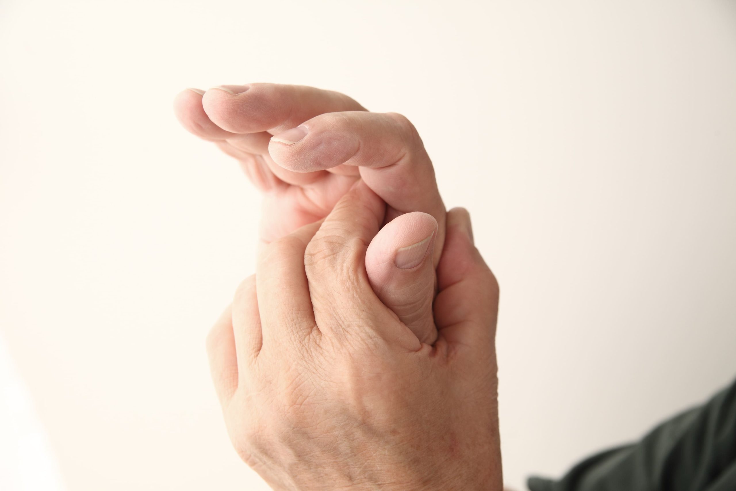
Immediate symptoms following UCL injury include pain at the inner aspect of the thumb base, swelling around the MCP joint, and difficulty with pinching. Bruising typically develops within 24-48 hours, often extending along the thumb’s inner border.
Pain Characteristics
Pain localises specifically to the ulnar (inner) side of the thumb MCP joint. Patients report sharp pain when attempting to grip objects or when the thumb is moved away from the hand. Direct pressure over the UCL insertion reproduces symptoms precisely.
Functional Limitations
Activities requiring pinch grip become notably difficult or painful. Patients often describe trouble with:
- Holding papers or cards firmly
- Turning doorknobs or keys
- Buttoning clothing
- Writing with a pen
- Gripping small objects between the thumb and index finger
Instability Signs
With complete tears, patients may notice the thumb “giving way” during gripping activities. Some describe a sense of looseness or weakness when attempting precision tasks. In severe cases, visible deviation of the thumb occurs during pinching attempts.
Clinical Examination and Diagnosis
Physical examination involves stress testing the UCL by applying an abduction force to the thumb whilst stabilising the metacarpal. Testing occurs in both extension and 30 degrees of flexion to assess both ligament components.
Stress Testing Interpretation
Increased laxity compared to the uninjured thumb suggests UCL damage. An endpoint, the sensation of the ligament stopping further movement, indicates a partial tear or sprain. Absence of an endpoint, particularly with laxity exceeding 30-35 degrees, suggests complete rupture.
Did You Know?
Local anaesthetic injection before stress testing may improve diagnostic accuracy by eliminating pain-induced muscle guarding that can mask true instability.
Imaging Studies
X-rays assess for avulsion fractures where the ligament pulls off a bone fragment. Avulsion fractures occur in a proportion of UCL injuries and influence treatment decisions.
Ultrasound can identify Stener lesions by visualising the displaced ligament position relative to the adductor aponeurosis. MRI provides detailed soft tissue assessment, showing ligament continuity, tear location, and Stener lesion presence with good accuracy.
Non-Surgical Management
Partial UCL tears with preserved joint stability heal effectively with immobilisation. A thumb spica splint or cast positions the thumb for optimal ligament healing whilst protecting against re-injury.
Immobilisation Protocol
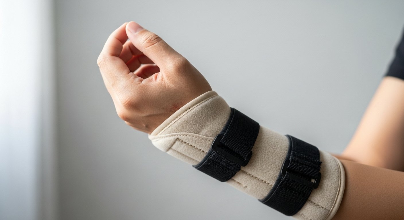
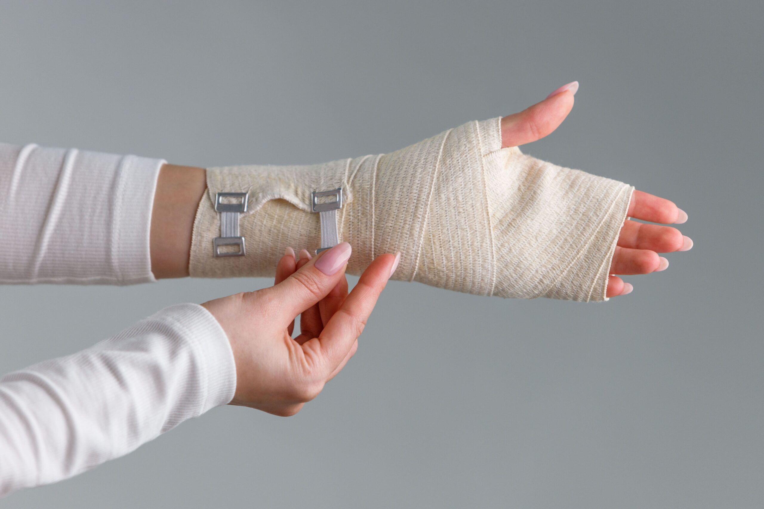
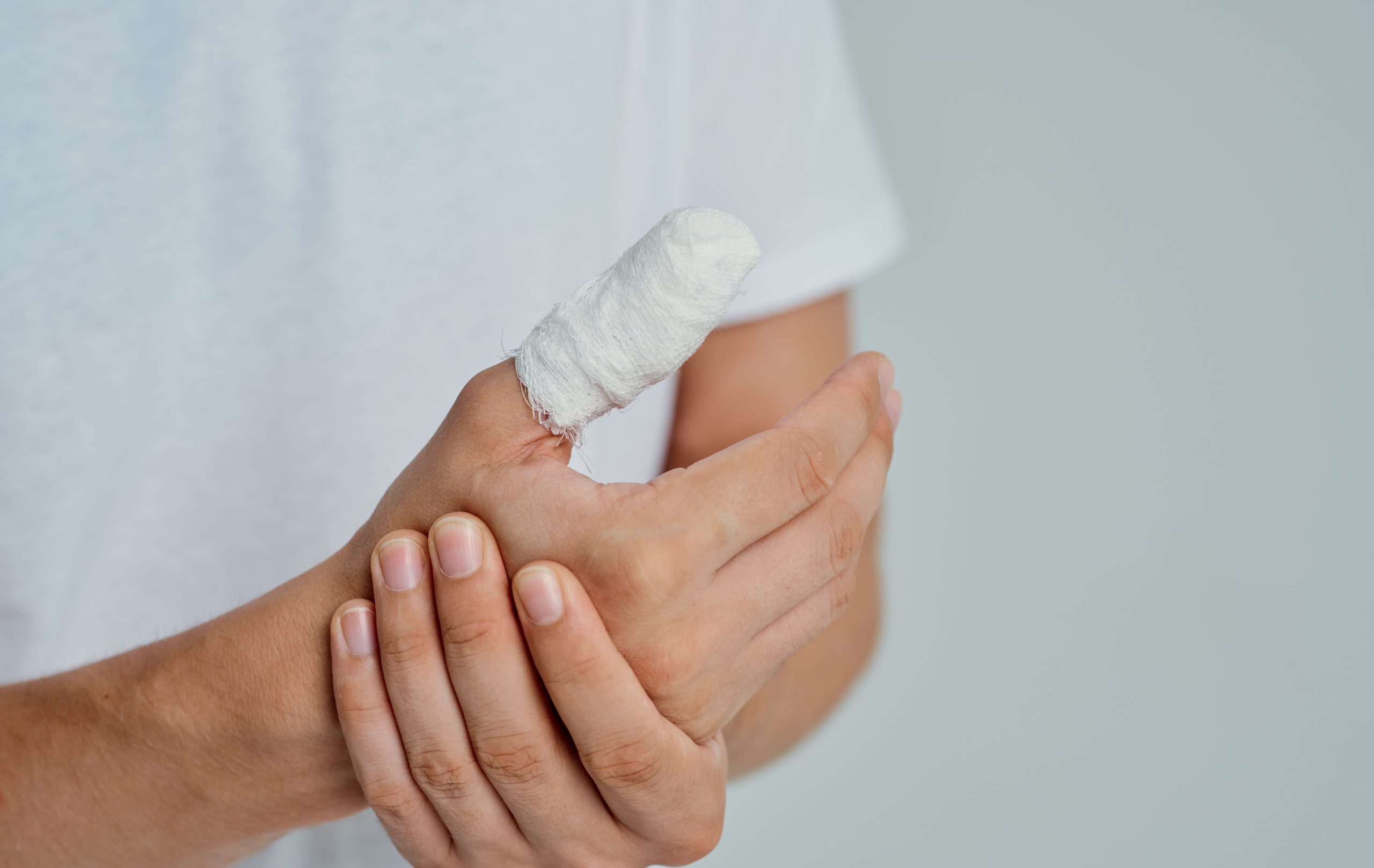
Treatment typically involves four to six weeks of continuous immobilisation in a thumb spica cast or splint. The MCP joint is positioned in slight flexion with the thumb in a functional position. The interphalangeal joint may be left free to maintain mobility.
Rehabilitation Phase
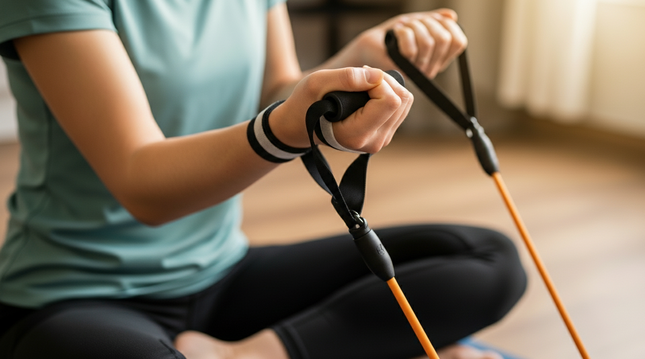
Following immobilisation, a graduated range of motion exercises restores thumb mobility. Strengthening begins once full motion returns, progressing from putty exercises to functional gripping activities. Return to sports or demanding manual activities typically occurs at ten to twelve weeks post-injury.
Expected Outcomes
Properly managed partial tears frequently achieve stable healing. Many patients regain functional grip strength and stability, though individual recovery depends significantly on compliance with the immobilisation timeline and rehabilitation program.
Surgical Repair Indications
Complete UCL tears, particularly with a Stener lesion, require surgical intervention for reliable healing. Other indications include:
- Joint instability exceeding 30-35 degrees on stress testing
- Displaced avulsion fractures
- Failed non-surgical treatment
- High-demand athletes or manual workers with complete tears
Timing Considerations
Early repair within three weeks of injury allows direct suture of the torn ligament to bone. Delayed presentation beyond six to eight weeks may find the ligament retracted and scarred, requiring reconstruction with a tendon graft rather than primary repair.
Important Note
Chronic UCL injuries presenting months after initial trauma typically require ligament reconstruction rather than repair, involving more extensive surgery and longer recovery.
Surgical Techniques
The specific procedure depends on injury pattern, timing, and tissue quality encountered during surgery.
Direct Ligament Repair
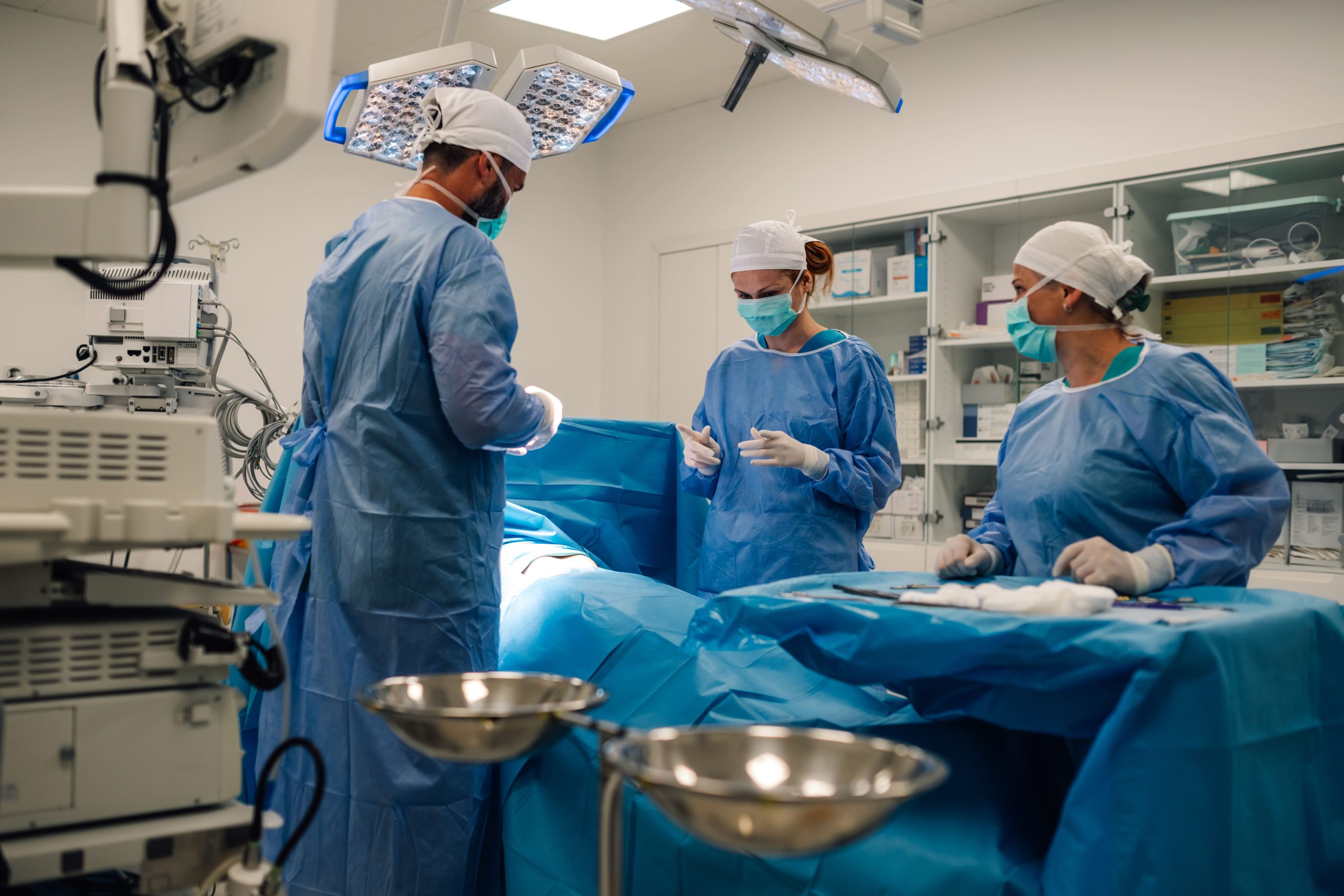
For acute injuries with adequate tissue quality, the surgeon reattaches the torn ligament to its anatomical insertion using suture anchors. These small devices embedded in bone provide secure fixation whilst the ligament heals. The adductor aponeurosis is repaired to restore normal anatomy.
Bone Anchor Fixation
Suture anchors allow secure ligament reattachment through small incisions. The anchor is placed at the ligament’s normal insertion site on the proximal phalanx, and sutures passed through the ligament are tied to restore anatomical tension.
Ligament Reconstruction
When primary repair is not possible due to chronic injury or poor tissue quality, reconstruction uses a tendon graft, typically palmaris longus from the forearm or a portion of an adjacent hand tendon. The graft recreates the ligament’s stabilising function through bone tunnels in the metacarpal and phalanx.
Post-Surgical Rehabilitation
Recovery protocols balance the protection of the repair against the need to restore motion and prevent stiffness.
Initial Protection Phase
A thumb spica splint or cast protects the repair for four to six weeks. During this period, gentle finger motion maintains tendon gliding whilst the ligament heals to the bone.
Motion Phase
Supervised hand therapy begins at four to six weeks with a gentle active range of motion. The therapist guides progression based on the healing assessment and individual response. Passive stretching is avoided initially to protect the repair.
Strengthening Phase
Progressive resistance exercises begin at eight to ten weeks. Activities include putty exercises, pinch strengthening, and a gradual return to functional tasks. Sport-specific or work-specific training follows once basic strength returns.
Return to Activities
Full activity clearance is commonly discussed at three to four months for non-contact activities and four to six months for contact sports or heavy manual work, subject to functional recovery. Some patients use protective taping during sports for the first season after surgery.
Clinical Outcomes
Clinical outcomes following timely skier’s thumb treatment are generally encouraging. Factors influencing results include accurate initial diagnosis, appropriate treatment selection based on injury severity, and patient compliance with rehabilitation protocols. Modern surgical techniques aim to restore mechanical stability to support functional hand recovery.
Optimising Your Recovery
- Follow immobilisation guidelines precisely. Premature removal of splints or casts risks re-injury or incomplete healing, potentially converting a successful non-surgical treatment into a surgical case.
- Attend all hand therapy sessions. Supervised rehabilitation ensures appropriate progression and identifies any complications early. Home exercises supplement but do not replace professional guidance.
- Protect the thumb during high-risk activities. Even after full recovery, taping or bracing during sports that originally caused injury provides additional security.
- Report new symptoms promptly. Increased pain, swelling, or instability during recovery warrants reassessment to ensure healing progresses appropriately.
When to Seek Professional Help
- Thumb pain and swelling following a fall or sports injury
- Difficulty gripping objects after thumb trauma
- Sensation of thumb instability or “giving way”
- Persistent thumb base pain lasting beyond several days
- Previous thumb injury with ongoing weakness or instability
- Bruising along the inner thumb border after injury
A comprehensive medical assessment can help distinguish a serious ligament tear from a mild thumb strain when symptoms persist after trauma.
Commonly Asked Questions
How can I tell if my thumb injury needs surgery?
The determining factors are joint stability on clinical examination and the presence of a Stener lesion on imaging. Complete tears with instability exceeding 30-35 degrees or a confirmed Stener lesion typically require surgical repair. A hand surgeon assesses these factors through physical examination and imaging studies to recommend an appropriate treatment pathway.
Will my grip strength return to normal after UCL repair?
Many patients regain functional grip strength following successful repair and structured rehabilitation. The recovery timeline varies among individuals, but meaningful clinical improvement is commonly seen within three months, with gradual functional gains continuing through six months.
What happens if I delay treatment for a UCL tear?
Delayed treatment may convert a straightforward primary repair into a more complex reconstruction. Over time, a torn ligament retracts and scars, losing the ability to be directly reattached to the bone. Reconstruction using a tendon graft can still achieve good functional results, but generally involves a longer recovery path.
Can skier’s thumb heal without surgery?
Partial tears with stable joints heal reliably with immobilisation alone. Complete tears without a Stener lesion may sometimes heal non-surgically, though clinical outcomes are less predictable than surgical stabilisation. Complete tears presenting with a Stener lesion cannot heal without surgery because the displaced tissue cannot contact its natural anatomical insertion point.
How long before I can return to sports after UCL surgery?
Participation in non-contact sports may resume around three to four months in suitable candidates. Contact sports and activities carrying a high risk of re-injury commonly require four to six months of healing and dedicated rehabilitation. Your surgeon and hand therapist guide return-to-sport clearance based on objective functional tracking.
Next Steps
Partial UCL tears with preserved joint stability typically respond well to immobilisation over four to six weeks. Complete tears, particularly those with a confirmed Stener lesion, generally require surgical repair within three weeks of injury for optimal clinical outcomes. Delayed presentation beyond this window may necessitate tendon graft reconstruction. Accurate diagnosis through stress testing and soft tissue imaging determines which treatment pathway applies.
If you are experiencing thumb pain, instability, or difficulty gripping following an injury, consulting a hand surgeon in Singapore can provide a comprehensive clinical evaluation to safely review your treatment options.